
Clinical Technique
Lip Anatomy, Aging, and a Practical Typology Framework for Filler Treatment Planning
A clinical guide to lip anatomy, aging changes, and a practical typology framework for aesthetic injectors planning filler treatments.
FAI Staff
FACE Aesthetic Institute
Lips are among the most technically demanding structures an aesthetic injector treats. The margin for error is narrow, the vascular anatomy is unforgiving, and patient expectations are often shaped by social media rather than anatomic reality. A confident, safe, and aesthetically consistent approach to lip augmentation starts with a clear understanding of the underlying anatomy, how that anatomy changes with age, and a repeatable framework for classifying what is sitting in front of you before the needle ever touches skin.
This article reviews perioral anatomy relevant to injection safety, summarizes the documented structural changes of lip aging, and outlines a practical typology approach that trainees and practicing injectors can use to organize assessment and treatment planning.
Perioral Anatomy Every Injector Should Know
The Orbicularis Oris and Philtral Columns
The orbicularis oris is a sphincteric muscle composed of two functionally distinct layers: the pars marginalis, which forms a continuous band running from modiolus to modiolus and governs fine lip movement and eversion, and the pars peripheralis, which lies more superficially and contributes to lip shape and posture. In the upper lip, fibers of the orbicularis oris decussate at the midline and insert into the dermis roughly 4 to 5 mm lateral to center, sparing the central philtrum. This insertion pattern pulls the overlying skin medially at two discrete points, producing the vertical ridges known as the philtral columns and the midline depression between them, the philtrum. The peaks where the columns meet the vermillion border form the Cupid's bow.
Understanding that the philtral columns are a muscular and dermal phenomenon, not simply a surface contour, matters clinically. Restoring definition to a flattened philtrum means restoring structural support along these insertion points, not just adding volume to the body of the lip.
The Vermillion Border and Wet-Dry Line
The vermillion border is the junction between the pigmented, keratinized red lip (vermillion) and the adjacent cutaneous (white) lip. In a youthful lip this border is crisp and well demarcated, which is part of what gives the lip its defined, three-dimensional appearance. The wet-dry line, or mucocutaneous line, is a separate landmark located posterior to the vermillion border, marking the transition between the dry (keratinized) vermillion and the wet, non-keratinized labial mucosa. Confusing these two landmarks is a common error among newer injectors, and it matters because of what runs beneath them.
Labial Artery Course and Danger Zones
The superior and inferior labial arteries, branches of the facial artery, supply the lips and run within or just deep to the submucosal plane, typically within or adjacent to the orbicularis oris, in the region posterior to the wet-dry line. Their depth and course are notably variable from patient to patient and can differ between the two sides of the same face, which means injectors should never assume a fixed, textbook location. In many patients the artery runs closer to the vermillion surface in the lateral thirds of the lip, increasing risk when injecting eversion technique near the commissures.
The clinical implication is straightforward: the area posterior to the wet-dry line, the philtral columns, and the central tubercle region are all higher-risk zones that warrant slow, low-volume, and ideally cannula-based or aspirating technique. Vascular occlusion in the lip can progress rapidly and, though rare, carries risk of tissue necrosis and, via retrograde arterial flow, more distant ischemic complications. Injectors should be fluent in recognizing early signs of vascular compromise and have a hyaluronidase protocol ready at every session.
How Lips Age: The Documented Changes
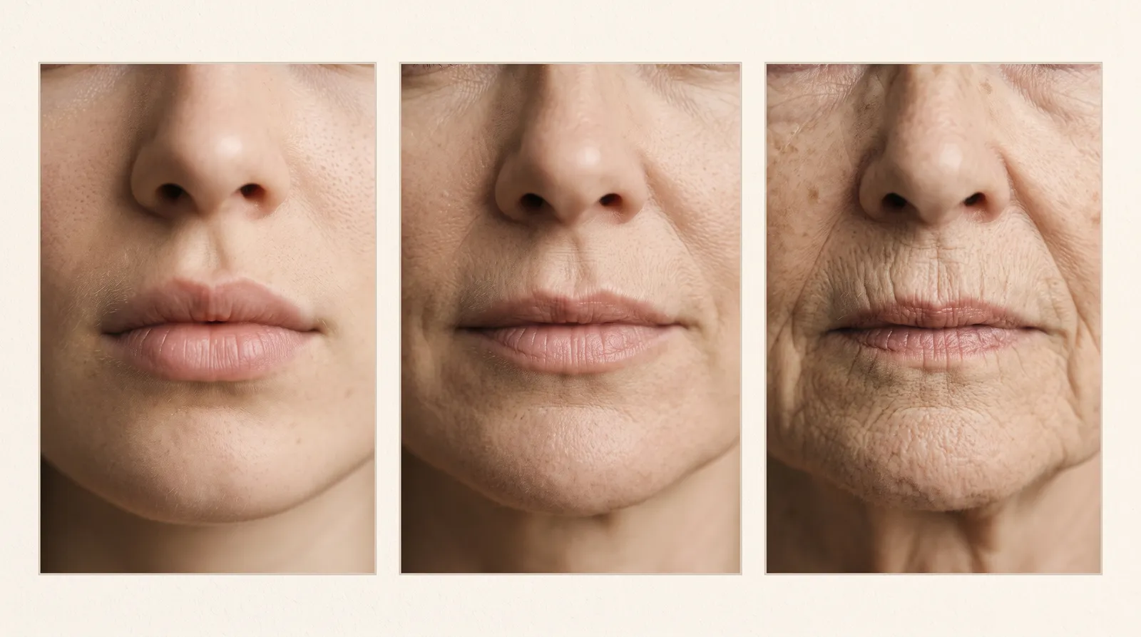
Lip aging is a composite process involving volume loss, dermal degradation, structural flattening, and altered proportions. It is not simply "lips getting thinner."
Volume Loss and Fat Compartment Changes
The lips and perioral region contain distinct superficial fat compartments that are thin from the outset and become progressively thinner with age. This is compounded by resorption of the maxilla and mandible, which reduces the bony scaffold that supports lip projection. As skeletal support recedes, the overlying soft tissue loses its anterior projection, and the lips appear to retrude and flatten even before significant skin laxity develops.
Dermal Degradation and Vermillion Effacement
Histologically, aging lip tissue shows a measurable decrease in dermal collagen and elastic fibers, along with thinning of the mucosal and cutaneous layers. This degradation reduces the structural crispness of the vermillion border, causing it to blur, or efface, into the surrounding cutaneous lip. The result is a lip that looks less defined in photographs and under makeup, independent of overall volume.
Philtral Column Flattening and Elongation
The dermal insertion points that create the philtral columns weaken with age, and the columns themselves flatten and become less distinct. At the same time, the vertical height of the philtrum increases substantially, with published measurements showing an increase from roughly 14 mm in youth to nearly 20 mm in older adulthood in female patients. This lengthening of the upper lip, combined with column flattening, is a major contributor to the "long upper lip" appearance common in mature faces and is a key reason why volume alone, injected without attention to vertical proportion, can make an aging lip look worse rather than better.
Loss of Definition and Postural Change
Additional hallmarks of the aging lip include blunting of the Cupid's bow, equalization of upper and lower lip projection (younger lips typically show a ratio favoring the lower lip), downturning of the oral commissures, and atrophy of the orbicularis oris itself, which reduces the muscle's contribution to lip eversion and pout. Perioral rhytids (radial lip lines) develop as the dermis thins and repetitive orbicularis contraction folds unsupported skin.
A Practical Typology Framework for Assessment
Rather than treating every patient with a single default injection pattern, experienced injectors benefit from categorizing lip presentations into recognizable types. This is not about forcing every patient into a rigid box. It is about training the eye to recognize dominant patterns so that technique, product selection, and volume can be matched to the actual anatomic deficit rather than applied generically.
Thin or Volume-Deficient Lips
Characterized by minimal vermillion show, a flat or absent tubercle, and reduced anterior projection, often in younger patients as a baseline anatomic trait rather than an aging change. Treatment planning generally prioritizes tubercle definition and border support over aggressive central bolus volume, to avoid an unnatural, overfilled look on a structurally thin frame.
Asymmetrical Lips
Present with visible differences in height, width, or projection between right and left halves, or between the two Cupid's bow peaks. Asymmetry may be congenital, post-traumatic, or related to dominant-side muscle use. Photographic documentation and precise mapping before injection are essential, since correcting asymmetry usually requires deliberately uneven product distribution.
M-Shaped or Gull-Wing Lips
Defined by an exaggerated, sharply angular Cupid's bow with pronounced central tubercles and comparatively flat or absent lateral tubercles, giving the upper lip an "M" or gull-wing silhouette. A published grading system classifies severity by the angle formed between the vertical midline through the central tubercle and the horizontal commissure line: mild at 15 degrees or less, moderate at 16 to 25 degrees, and severe above 25 degrees. Treatment for more severe presentations often focuses on building out the underdeveloped lateral tubercles to soften the angularity, rather than adding further volume to an already prominent central peak.
Septated or Multi-Tubercle Lips
Lips displaying more than the typical three tubercles (two lateral, one central) or unusual segmentation across the vermillion body. This variant is addressed within tubercle-based classification systems, which organize treatment around the number, distribution, and prominence of tubercles present, rather than treating the lip as a single undifferentiated unit.
Mature or Aged Lips
Show the composite aging pattern described above: flattened philtral columns, effaced vermillion border, elongated white lip, reduced pout, downturned commissures, and often perioral rhytids. These patients typically need a layered approach addressing structural support (vermillion border and philtral columns), volume restoration, and sometimes adjunctive treatment of perioral rhytids and commissure descent, rather than central body volume alone.
Applying the Framework to Treatment Planning
A structured assessment sequence, patient interview and goals, static and dynamic examination, anatomic and vascular risk assessment, typology classification, then technique selection, produces more consistent, natural results than an intuitive or purely volumetric approach. Recognizing which typology (or combination of typologies) is present allows the injector to choose appropriate vectors, prioritize border restoration versus bulk volume, and set realistic expectations with the patient before treatment begins.
Conclusion
Lips age through a combination of skeletal resorption, fat compartment thinning, dermal collagen loss, and muscular atrophy, changes that manifest as flattened philtral columns, an effaced vermillion border, elongated upper lip height, and reduced projection. Layering a typology-based assessment on top of this anatomic knowledge gives injectors a repeatable, defensible framework for treatment planning that respects each patient's baseline structure rather than applying a one-size-fits-all injection pattern.
Sources
- Anatomical-Based Diagnosis and Filler Injection Techniques: Lips and Philtrum. PMC (National Library of Medicine).
- Navigating the Labial Artery: A Safer Approach to Submucosal Lip Filler Techniques. PMC (National Library of Medicine).
- Innovative Classification of M-Shaped Lips for Enhanced Refill Outcomes: A Case Series. Journal of Cosmetic Dermatology, via PMC.
- Introduction of the Lip Classification of Tubercles: A Novel Approach to Minimally Invasive Aesthetic Lip Treatments. PMC (National Library of Medicine).
- Universal Lip Integrity, Proportion, and Structure (uLIPS): A Framework for Achieving Consistent, Natural-Looking Lip Augmentation Using Hyaluronic Acid Fillers. PMC (National Library of Medicine).
- Lips and Perioral Region Anatomy. Medscape Reference (Emedicine).
Disclaimer
This article is intended for the professional education of licensed aesthetic providers and students in training. It is not a substitute for hands-on clinical instruction, supervised injection practice, or the individualized clinical judgment required in direct patient care. All injectors should pursue accredited hands-on training and operate within their scope of practice and local regulatory requirements.
Continue reading
More articles.

Clinical Technique
Recognizing and Managing Vascular Occlusion in Dermal Filler Injection
A clinical guide for injectors on recognizing filler-induced vascular occlusion, danger zones, warning signs, and emergency hyaluronidase protocols.
FAI Staff9 min
Clinical Technique
Why Skin Quality Belongs in Every Injectable Consultation
Why skin quality assessment belongs in every injectable consult, and how to sequence skincare, biostimulators, and devices with toxin and filler.
FAI Staff8 min
Clinical Technique
Laser Physics Fundamentals and Chromophore Targeting: A Clinical Primer
Learn selective photothermolysis and chromophore targeting (melanin, hemoglobin, water) to guide laser device selection for aesthetic providers.
FAI Staff9 min
Clinical Technique
HA vs. Non-HA Dermal Fillers: How to Choose the Right Product for the Right Goal
A clinician's guide to choosing HA versus biostimulatory fillers like Radiesse, Sculptra, and Bellafill by goal, area, and reversibility.
FAI Staff9 min