
Clinical Technique
Facial Fat Compartments: Why Anatomy Should Drive Every Filler Plan
A clinical overview of facial fat compartment anatomy and how it should guide filler placement, volume assessment, and injection planning.
FAI Staff
FACE Aesthetic Institute
Facial aging is often described as a global process of sagging and thinning. That description is convenient, but it is not anatomically accurate. The face does not lose volume evenly, and it does not age as a single sheet of tissue sliding downward.
Instead, the subcutaneous and deep fat of the face is organized into discrete, fascially bound compartments, each with its own blood supply, its own rate of age-related change, and its own relationship to the structures beneath it. For injectors, understanding this compartmentalized anatomy is not academic detail. It is the foundation of safe, natural-looking, and reproducible volumizing treatment.
The Compartmentalized Face: A Shift in Understanding
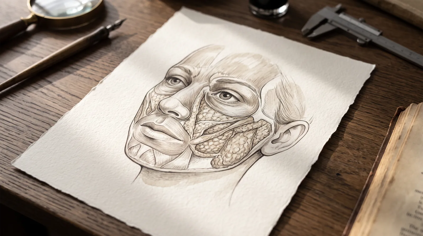
For much of the history of facial aesthetic medicine, the subcutaneous fat of the face was treated as one continuous layer that simply thinned and drooped with age. That model changed substantially following the cadaveric dissection work of Rohrich and Pessa in the mid-2000s, which used dye injection studies to map the boundaries of facial fat and demonstrated something clinically important: facial fat is not continuous. It is divided into separate anatomic units, separated by fibrous septa, each functioning as an independent compartment.
This work, along with subsequent anatomic studies, established that the face contains both superficial and deep fat compartments, arranged in layers relative to the superficial musculoaponeurotic system (SMAS) and the deeper facial mimetic muscles. Each compartment has its own vascular pedicle, which is part of why volume loss, fat grafting outcomes, and even complication patterns vary so much by location.
Superficial Fat Compartments
The superficial fat lies above the SMAS and is organized into named units separated by retaining ligaments and fascial septa. In the midface and cheek, these include the nasolabial fat compartment, the superficial medial cheek fat, the middle cheek fat, and the lateral temporal cheek fat. The forehead has its own central, middle, and lateral temporal-cheek subdivisions, and the lower face includes the jowl fat compartment, the most inferior of the subcutaneous units.
The periorbital region has its own superficial orbital fat compartments, bounded by septal attachments, distinct from the deeper orbital fat that contributes to true fat pad prolapse.
Deep Fat Compartments
Beneath the SMAS lies a separate set of compartments that sit directly on, or just superficial to, the facial skeleton and the mimetic muscles. In the midface, the key deep compartments are the deep medial cheek fat (DMCF), often described as having a medial and lateral component, and the suborbicularis oculi fat (SOOF), which is likewise divided into medial and lateral portions. The buccal fat pad, with its extensions, also occupies a deep plane in the lower midface.
The deep medial cheek fat is particularly important clinically. It sits against the maxilla, close to the piriform aperture, and functions as the structural foundation that projects and supports the overlying superficial malar fat. Loss of volume here has an outsized effect on midface appearance relative to its actual size.
Why Volume Loss Is Not Uniform
If facial fat were a single continuous layer, aging would simply flatten it evenly. Because the face is compartmentalized, aging instead produces a patchwork of change: some compartments atrophy substantially, some are relatively preserved, and a few may even expand.
Longitudinal imaging studies following patients over roughly a decade have shown that deep fat compartments lose volume disproportionately compared to superficial compartments, with deep fat volume decreasing by around 18 percent versus roughly 11 percent in superficial fat over a similar interval. The deep medial cheek fat and the SOOF are frequently cited as compartments especially prone to early, significant atrophy. Meanwhile, some superficial compartments, particularly around the jowl and nasolabial region, can appear relatively preserved or even prominent by comparison, not because they are gaining volume, but because the deep support beneath them has been lost.
This differential atrophy is the anatomic explanation for several classic signs of facial aging:
- Loss of deep medial cheek fat and SOOF contributes to a flattened, deflated midface and a visible step-off at the lid-cheek junction, often called the "tear trough" or V-deformity.
- Descent and relative prominence of the nasolabial fat compartment deepens the nasolabial fold, not primarily because that compartment enlarges, but because it loses its deep structural support and settles inferiorly.
- Jowl fat, unrestrained by the loss of midface support above it, becomes more visually apparent along the mandibular border.
- Pseudoherniation of orbital fat in the lower eyelid occurs as septal attenuation allows deeper orbital fat to bulge forward, a distinct process from cheek compartment atrophy but often discussed alongside it.
Pseudoptosis: Descent Without True Sagging
One of the most clinically useful concepts to come from compartment anatomy is pseudoptosis: the appearance of tissue "sagging" that is actually caused by loss of underlying volume rather than by skin laxity or gravity alone.
When the deep medial cheek fat and SOOF atrophy, the superficial fat compartments that rested on top of them lose their structural scaffold. The superficial tissue does not necessarily stretch or descend on its own; it settles downward and inward because the foundation beneath it has deflated. Clinically, this distinction matters enormously. A face with significant pseudoptosis from deep volume loss may respond very well to deep compartment volumization, while a face with true skin and ligamentous laxity may need a different treatment strategy altogether, or a surgical referral.
Failing to distinguish between these mechanisms is one of the more common planning errors in injectable practice. Treating pseudoptosis as though it were purely a skin-laxity problem, or vice versa, leads to mismatched expectations and suboptimal outcomes.
Why This Anatomy Should Drive Filler Planning
A compartment-based approach to volumizing treatment starts with a simple premise: restore volume in the plane and location where it was actually lost, rather than adding volume superficially wherever a fold or hollow is visible.
Treating the Deep Plane First
Because deep compartments like the DMCF and SOOF function as structural support, many experienced injectors prioritize restoring volume in the deep plane before addressing superficial folds. Placing product at or near the periosteal or supraperiosteal level in the deep medial cheek fat can restore anterior projection to the midface and indirectly soften the nasolabial fold and lid-cheek junction, without ever injecting into those areas directly. This is sometimes referred to as building a supportive "pillar" beneath the superficial tissue.
This approach also tends to produce a more natural result because it works with the face's own architecture rather than against it. Injecting superficially and centrally to camouflage a fold that is actually caused by deep volume loss can create a puffy, overfilled appearance without correcting the underlying structural deficit.
Matching Product and Technique to the Compartment
Different compartments favor different injection depths, tools, and product rheology. Deep compartments, positioned close to the periosteum, are typically treated with a stiffer, higher-G-prime filler placed via needle or cannula in small aliquots or boluses to rebuild projection. Superficial compartments generally call for softer products and more conservative volumes, since they sit closer to the skin surface where firmness or lumpiness is more visible and where vascular structures may be more superficial and mobile.
Using a deep, structural product superficially, or a soft dynamic product where structural support is needed, is a mismatch that can compromise both safety and aesthetic outcome.
Avoiding Overfilling and Wrong-Plane Injection
Overfilling is often less about total volume and more about placement. Injectors who do not think in terms of compartments may repeatedly add product to the same superficial area that looks deficient, without recognizing that the actual deficiency lies in a deeper compartment. The result is bulk without the correct contour, an appearance many patients and providers now recognize as an "overfilled" look.
Compartment anatomy also carries direct safety implications. Knowledge of which compartments lie adjacent to major vascular structures, such as the facial artery and its branches as they course near the nasolabial fold and deep medial cheek fat, or the angular artery near the piriform region, allows injectors to select safer entry points, injection depths, and techniques (including cannula use where appropriate) to reduce the risk of intravascular injection and its complications.
Clinical Takeaway
Facial volume loss is not a uniform process, and effective, safe filler treatment cannot be planned as though it were. Deep fat compartments, particularly the deep medial cheek fat and the suborbicularis oculi fat, tend to atrophy earlier and more significantly than many superficial compartments, and this differential loss is what drives much of the visible change associated with an aging midface.
A compartment-based framework for assessment and treatment planning, evaluating which specific fat compartments have lost volume, in which plane, and how that loss is affecting the compartments around it, allows injectors to restore structure rather than simply disguise folds. This produces results that look proportionate and natural, and it reduces the risk of overfilling, wrong-plane injection, and avoidable vascular complications.
Sources
- Rohrich, R.J., Pessa, J.E. "The Fat Compartments of the Face: Anatomy and Clinical Implications for Cosmetic Surgery." Plastic and Reconstructive Surgery, 2007.
- "The Clinical Importance of the Fat Compartments in Midfacial Aging." Plastic and Reconstructive Surgery.
- "Facial Fat Compartments: A Guide to Filler Placement." PMC (PubMed Central).
- "Facial fillers: Relevant anatomy, injection techniques, and complications." PMC (PubMed Central).
- "Injection Guidelines for Treating Midface Volume Deficiency With Hyaluronic Acid Fillers: The ATP Approach (Anatomy, Techniques, Products)." Aesthetic Surgery Journal, Oxford Academic.
- American Society for Dermatologic Surgery / American Academy of Facial Plastic and Reconstructive Surgery: professional resources on facial anatomy and injectable safety education for clinicians.
Educational Disclaimer
This article is intended for professional education purposes for licensed aesthetic providers and students in training. It is not a substitute for hands-on clinical instruction, cadaver-based anatomy training, or supervised injectable practice under qualified proctorship. Injectable treatment planning should always be individualized to the patient and performed within the scope of the provider's training, licensure, and applicable regulations.
Continue reading
More articles.

Clinical Technique
Recognizing and Managing Vascular Occlusion in Dermal Filler Injection
A clinical guide for injectors on recognizing filler-induced vascular occlusion, danger zones, warning signs, and emergency hyaluronidase protocols.
FAI Staff9 min
Clinical Technique
Why Skin Quality Belongs in Every Injectable Consultation
Why skin quality assessment belongs in every injectable consult, and how to sequence skincare, biostimulators, and devices with toxin and filler.
FAI Staff8 min
Clinical Technique
Laser Physics Fundamentals and Chromophore Targeting: A Clinical Primer
Learn selective photothermolysis and chromophore targeting (melanin, hemoglobin, water) to guide laser device selection for aesthetic providers.
FAI Staff9 min
Clinical Technique
Lip Anatomy, Aging, and a Practical Typology Framework for Filler Treatment Planning
A clinical guide to lip anatomy, aging changes, and a practical typology framework for aesthetic injectors planning filler treatments.
FAI Staff8 min